Ultrasound
Accredited by the American College of Radiology
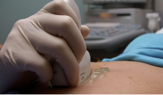
Ultrasound uses high frequency sound waves to visualize soft tissue organs and vascular structures. It is a painless non invasive way to evaluate a number of internal organs.
General & OB Exams
OB/Pregnancy Ultrasound
Uses high frequency sound waves to visualize an embryo or fetus. We evaluate pregnancy, fetal development, and due dates. We offer 4-D images and are certified by The Nuchal Translucency Quality Review Program.
Abdominal & Organ
Pelvic, Abdominal, Gallbladder, Kidney, Bladder, Pancreas and Scrotum Ultrasound. Painless visualization of internal organs.
Thyroid & Neck
Evaluation of soft tissue structures of the neck, including the thyroid gland.
Vascular (Doppler)
Specialized imaging to view blood flow in veins or arteries. Used to identify blood clots, aortic aneurysms (AAA screening), or plaque in carotid arteries.
Sonohysterogram
Visualizes the endometrium using a probe and catheter. Procedure takes approx 20-30 minutes.
Thoracentesis / Paracentesis
Ultrasound-guided procedures to locate and drain fluid from the chest or abdomen.
Breast Procedures
Breast Ultrasound
Often performed as a supplement to mammography using high frequency sound waves for high-resolution images.
Ultrasound Guided Breast Biopsy
The radiologist locates tissue, anesthetizes the area, and takes samples with a vacuum-assisted needle. A small marker is placed, and a mammogram follows to confirm placement.
Breast Aspiration
Under ultrasound guidance, fluid is removed from a cyst or abnormal area using a small needle. Fluid may be sent to a lab.
Vero Radiology Women's Imaging Center is fully accredited by the American College of Radiology.
Ultrasound Exam Prep
General Ultrasound Prep
- OB/GYN (Pelvic) & Bladder only: Drink 32 oz of water one hour prior to exam. Finish water one hour before. Do Not Urinate prior to exam. (Patients <16 yrs contact office; >24 weeks pregnant do not need to drink water).
- Gallbladder / Abdomen / Aorta / Kidney: No food, drink, or medication 6-8 hours prior.
- Kidney only (Renal): No Prep required.
Biopsy & Aspiration Prep
- Discontinue aspirin 7 days prior.
- Discontinue Vitamin E, Plavix, and anticoagulants 5 days prior.
- Discontinue blood thinners (Coumadin) 4 days prior (PT, PTT, INR labs needed 24 hours prior).
- Discontinue Gingko Biloba 2-3 days prior.
- Please contact your physician prior to discontinuing any medications.